Fast Fluorescence Imaging using Radiofrequency-tagged Emission
New development in microscopy inspired by wireless technology eliminates the speed limitation in biological imaging for blood diagnostics and brain mapping
Fluorescence imaging is the most widely used method to analyze the molecular composition of biological specimens. Target molecules, when they are present, can be “tagged” with a fluorescence label and made visible. For example, the technique is used in screening blood for cancer cells, or to study biochemical reactions. In particular, the highly sensitive imaging technique is very good at detecting molecules present at extremely low concentrations.
However, because of the very small amount of light produced by fluorescent molecules, current state-of-the-art camera technologies must image samples slowly, in order to collect enough photons to generate an image. Further, these cameras typically read out individual pixels one at a time to create an image. These two factors limit the speed at which fluorescence imaging can be performed.
Inspired by how wireless communication networks uses multiple radio frequencies to communicate with multiple users, and our own past work on radio frequency photonics, the Jalali-Lab has developed a new high-speed microscopy technique that is an order of magnitude faster than current fluorescence imaging technologies.
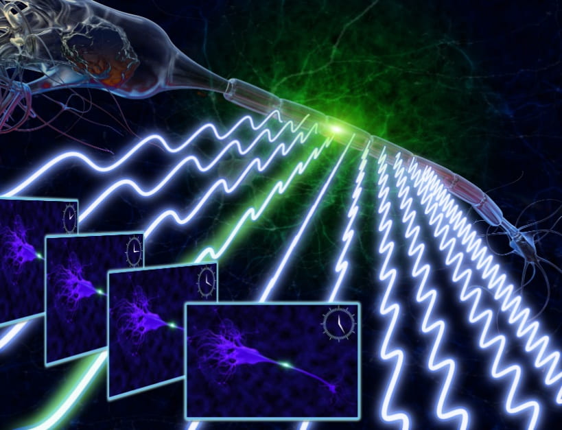
Our camera encodes fluorescence emission from each pixel on a different radio frequency (RF). Each pixel on the image functions as a different RF channel. The light from all pixels is collected using a single pixel photodetector. The RF spectrum obtained via digital Fourier transform reveals the fluorescent image. The “fluorescence imaging using radiofrequency-tagged emission”, or FIRE, is about 10 times faster than other techniques.
Due to the high imaging speed, the technique can be applied in flow cytometry – which analyzes cells one-by-one in a flowing fluid. Unlike current imaging flow cytometers, the FIRE technique can keep up with conventional flow cytometry speeds, and image 50,000 cells per second. While most flow cytometers take only a single-point estimate of a cell’s fluorescence, FIRE can create an entire high-resolution, multi-color image of each cell, which can provide significantly more information to a biologist or clinician. This allows us to both detect and differentiate rare cells at high throughputs. The new technique can also be applied in vivo, for example, in neuroscience research to view neuron activity in the brain. Here the sub-millisecond response of the camera will image the action potential activity of neurons.
We have extended radio frequency tagging idea to create a new instrument for dynamic light scattering (Mie scattering) for characterization of particles such as biological cells or pollutants in air and water. Ratio frequency encoded angular light scattering (REALS) measures the angular spread of scattered light in single shot with a single photodetector and without the need for mechanical scanning. It does so by encoding the optical excitation angle to radio frequency and obtains the angular spectrum of scattered light by simply examining the RF spectrum of scattered light measured at a single angle. Furthermore, the radio frequency encoding has led to a new method for precision measurement the fluorescent lifetime.
-
Brandon W. Buckley, Najva Akbari, Eric D. Diebold, Jost Adam, and Bahram Jalali, “Radiofrequency encoded angular light spectroscopy”, Applied Physics Letters, (2015). link pdf
-
J. C.K. Chan et al., “Digitally synthesized beat frequency multiplexed fluorescence lifetime spectroscopy”, Biomedical Optics Letters, (2014). link pdf
-
Eric D. Diebold, BrandonW. Buckley, Daniel R. Gossett and Bahram Jalali, “Digitally synthesized beat frequency multiplexing for sub-millisecond fluorescence microscopy,” Nature Photonics, (2013).link pdf”
-
Researchers develop new type of fluorescent camera for blood diagnostics, brain mapping”, Phys.Org. link


